Microscopic Gallery
Original observation, recorded as it was seen.
Every plate below is an original photograph from the founder's own gemological work. Captions describe what was observed and nothing beyond it — no identity, origin or treatment is asserted without established evidence.
From My Gemological Archive
Real specimens. Real observations. Original imagery from my gemological work.
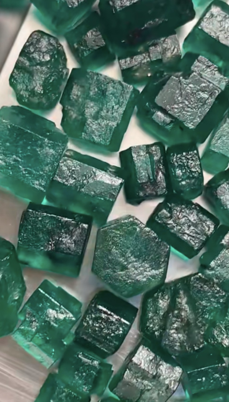 Plate 004
Rough Emerald
Rough emerald crystals documented for visual record and morphological comparison. The observation is limited to visible crystal form, surface features and colour, without adding claims regarding treatment or origin.
From the EGP Gemological Archive — Photograph by Eman Galeb Hamza
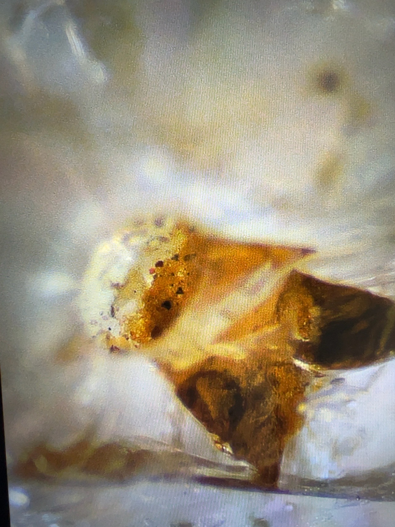 Plate 005
Longitudinal Yellow Inclusion in Treated Sapphire
An elongated yellow internal inclusion in a treated sapphire specimen, documented as a longitudinal morphological feature under the recorded microscopic illumination. The specific treatment type or inclusion composition is not inferred from appearance alone.
From the EGP Gemological Archive — Photograph by Eman Galeb Hamza
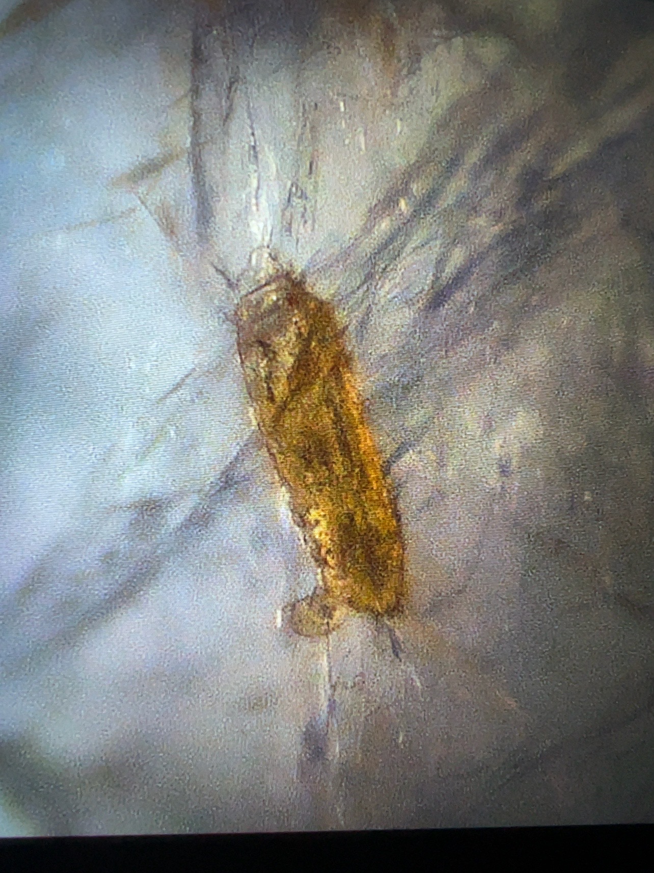 Plate 006
Bleeding-like Inclusion in Treated Ruby
An internal inclusion in a treated ruby specimen showing an appearance described as bleeding-like, which is a purely visual description. It does not imply biological bleeding, nor does the form infer a specific treatment mechanism.
From the EGP Gemological Archive — Photograph by Eman Galeb Hamza
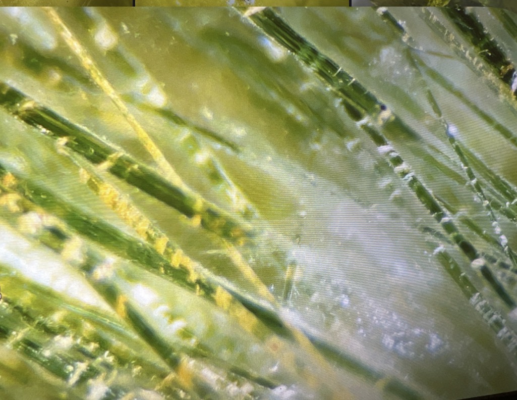 Plate 007
Green Rutile Inclusions in Quartz
General microscopic view showing a group of elongated green rutile crystals enclosed within quartz, with a clearly visible linear distribution of the inclusions in the host.
From the EGP Gemological Archive — Photograph by Eman Galeb Hamza
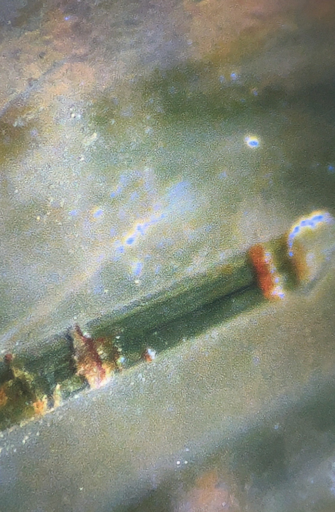 Plate 008
Microscopic Detail of Green Rutile in Quartz
Closer microscopic detail showing an elongated green rutile crystal within quartz, highlighting the acicular form and the crystalline boundaries of the internal inclusion.
From the EGP Gemological Archive — Photograph by Eman Galeb Hamza
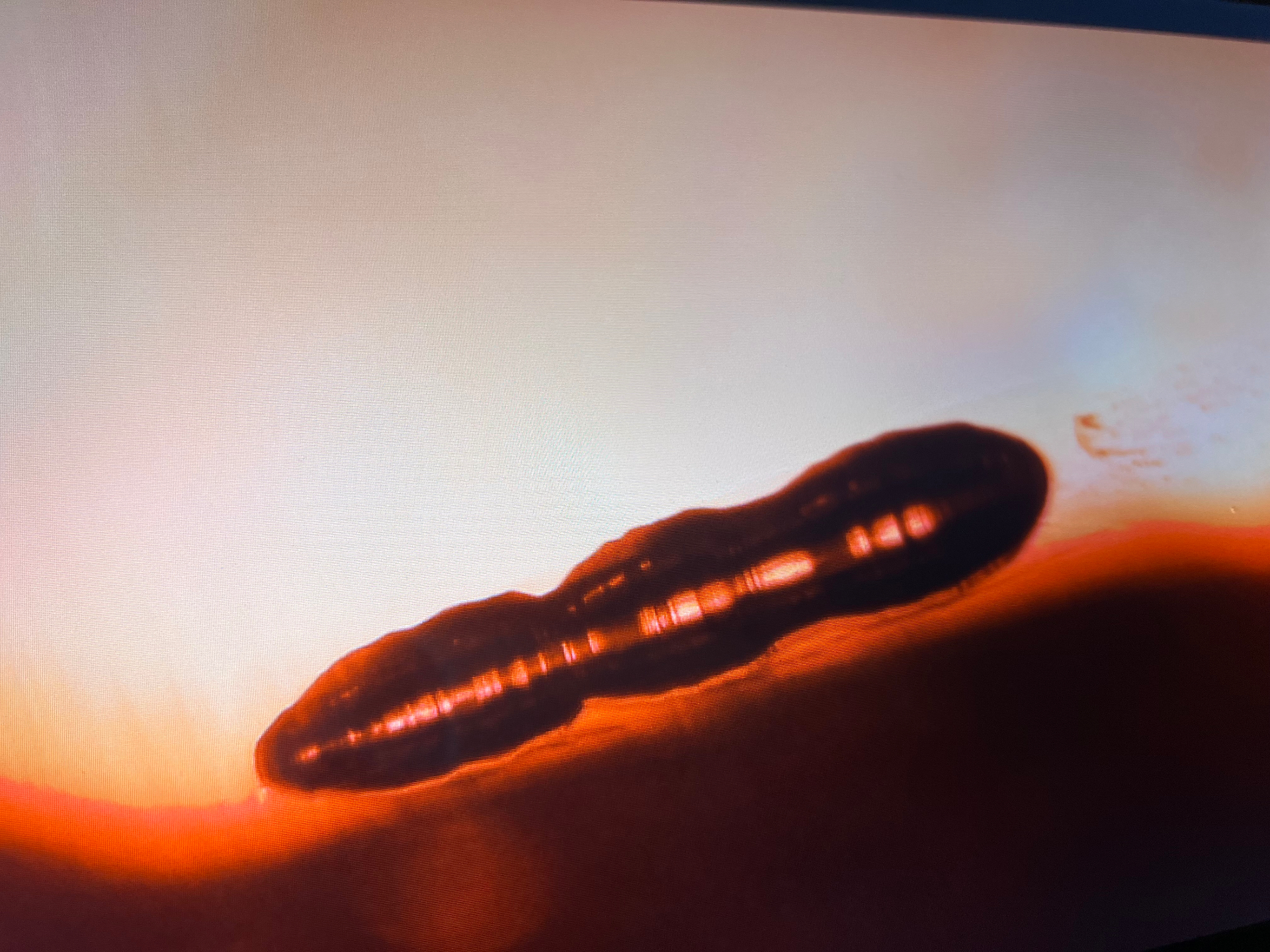 Plate 009
Elongated Tubular Internal Feature in Synthetic Ruby
Microscopic view of a synthetic ruby showing an elongated tubular internal feature, with its morphology clearly visible under transmitted light during gemological examination. The feature is documented as an internal characteristic of the specimen without assigning a specific formation mechanism from visual evidence alone.
From the EGP Gemological Archive — Photograph by Eman Galeb Hamza
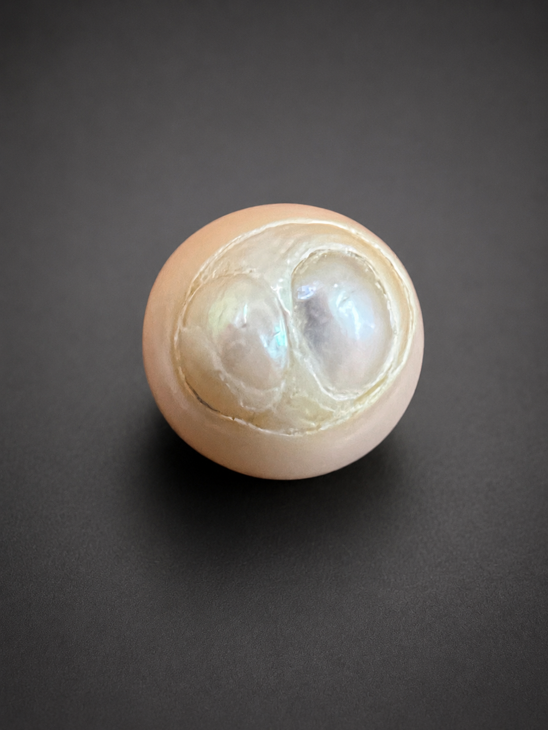 Plate 010
Twin Pearl — Internal Structural View
A pearl showing a distinct twin internal structure, with two adjacent pearl bodies developed within a single host, revealing nacre layering and internal growth architecture.
From the EGP Gemological Archive — Photograph by Eman Galeb Hamza
Observation Series
Graphite-Rich Specimen Observation Series
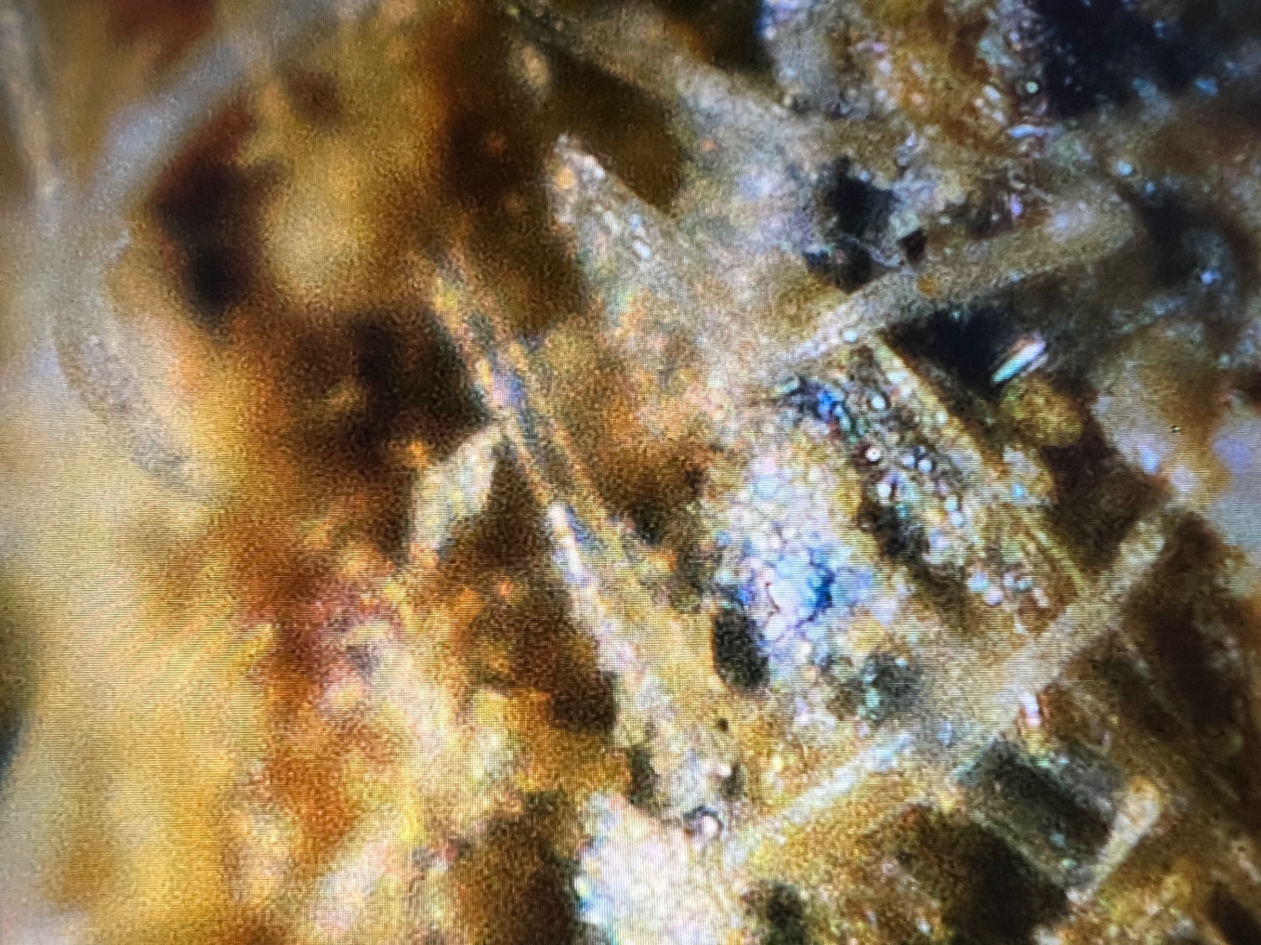 Plate 011
Microscopic Detail of a Graphitic Section Associated with Untreated Rough Diamond
Closer microscopic view of a prepared section taken from the graphite-rich material that surrounded an untreated rough diamond crystal. The section reveals fine elongated tubular and linear internal features together with heterogeneous dark areas. These features are documented morphologically as observed under magnification, without assigning a specific mineralogical composition or formation mechanism from visual evidence alone.
Specimen observation: the associated rough diamond crystal showed unusually high resistance and notable difficulty during cutting. This is recorded as an independent practical observation and is not presented as evidence of a causal relationship with the microscopic features shown.
From the EGP Gemological Archive — Photography and documentation by Eman Galeb Hamza
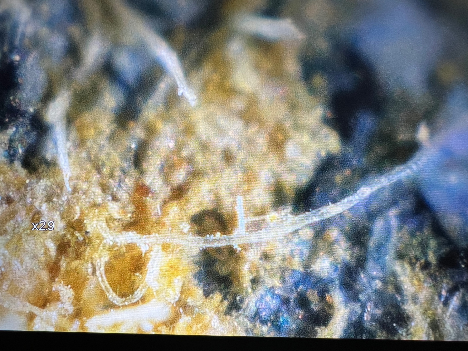 Plate 012
Overview of a Graphitic Section Formerly Surrounding a Rough Diamond Crystal
Lower-magnification microscopic overview of a section taken from the graphite-rich layer that surrounded an untreated rough diamond crystal. The field shows a network of elongated linear and tubular features distributed through the material, documenting their spatial distribution and textural relationships before closer microscopic examination.
From the EGP Gemological Archive — Photography and documentation by Eman Galeb Hamza